
The CBCT knows where the bone and the nerve are. It does not know what the tooth surfaces look like — not at the fidelity a guide has to seat on. That is what the optical scan is for, and loading it is a step of its own before the two are brought into the same coordinate system.
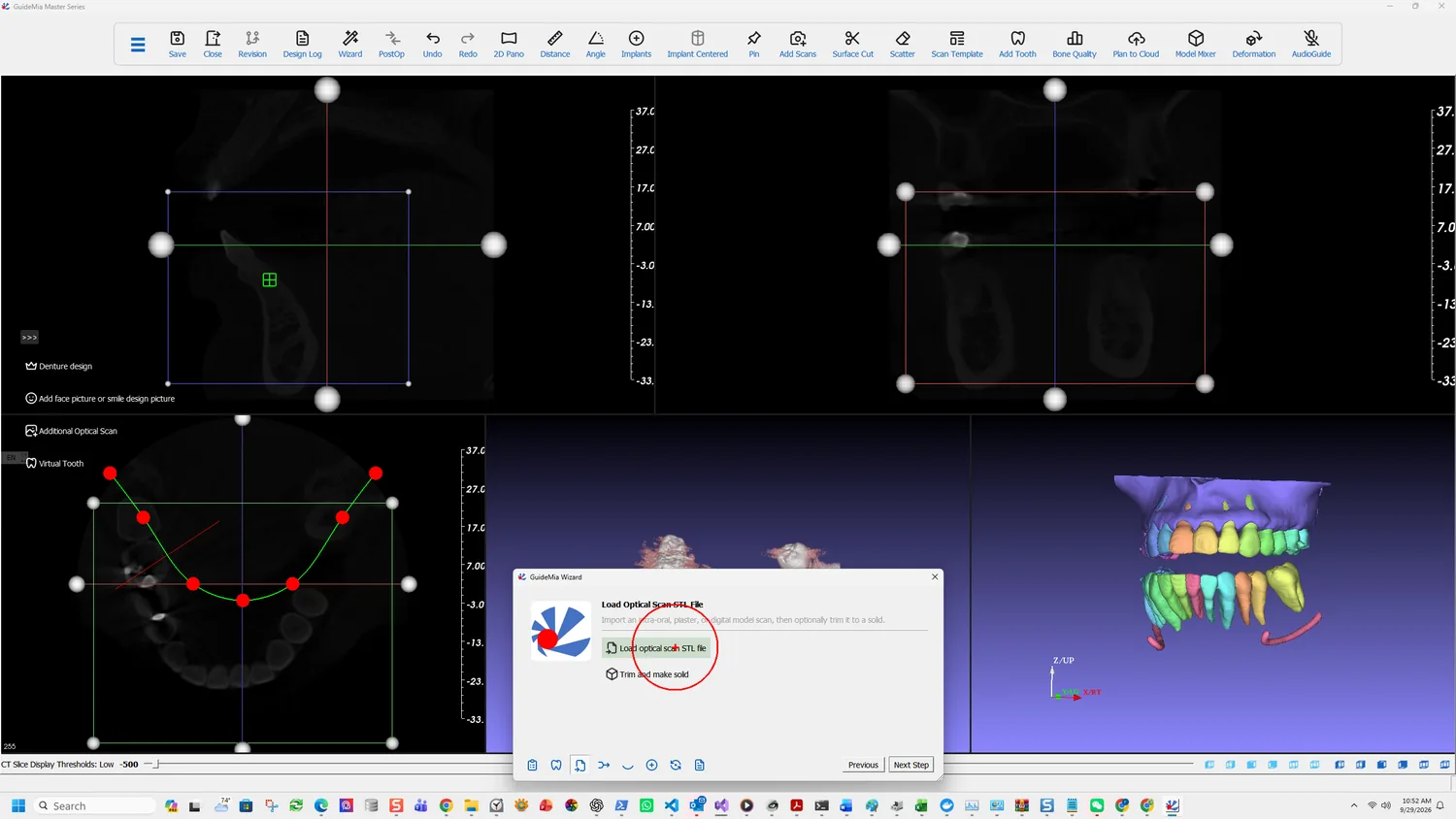
What counts as an optical scan
The software is deliberately unfussy about where the surface data came from:
Load optical scan of the teeth and gingiva. You can use a plaster model scan, an intra-oral scan, or even a digital model designed from another system. This is for partially edentulous cases so that surgical guides will be generated using the scan file.
Three routes, then: an intraoral scan, a scan of a plaster model, or a digital model built elsewhere. All arrive as a mesh file and all serve the same purpose.
The last sentence states the indication plainly. This is the partially edentulous route — the guide will be generated from the scan, seating on teeth that exist. A fully edentulous case has no such surfaces and goes the radiographic guide route instead.
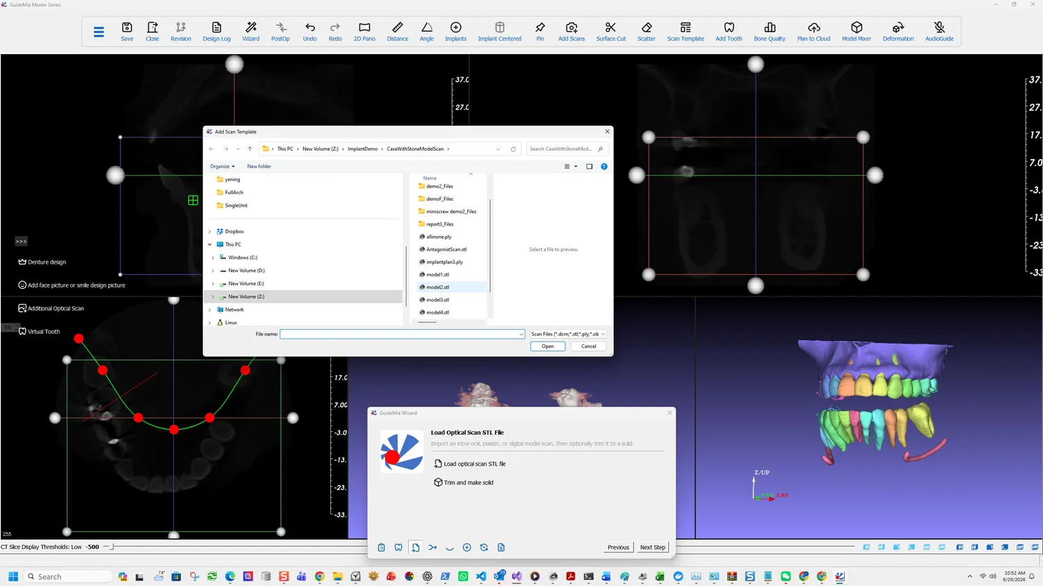
Add Scan Template opens an ordinary file dialog. The scan loads as its own object, in its own coordinates, and sits alongside the CT-derived models without any relationship to them yet.
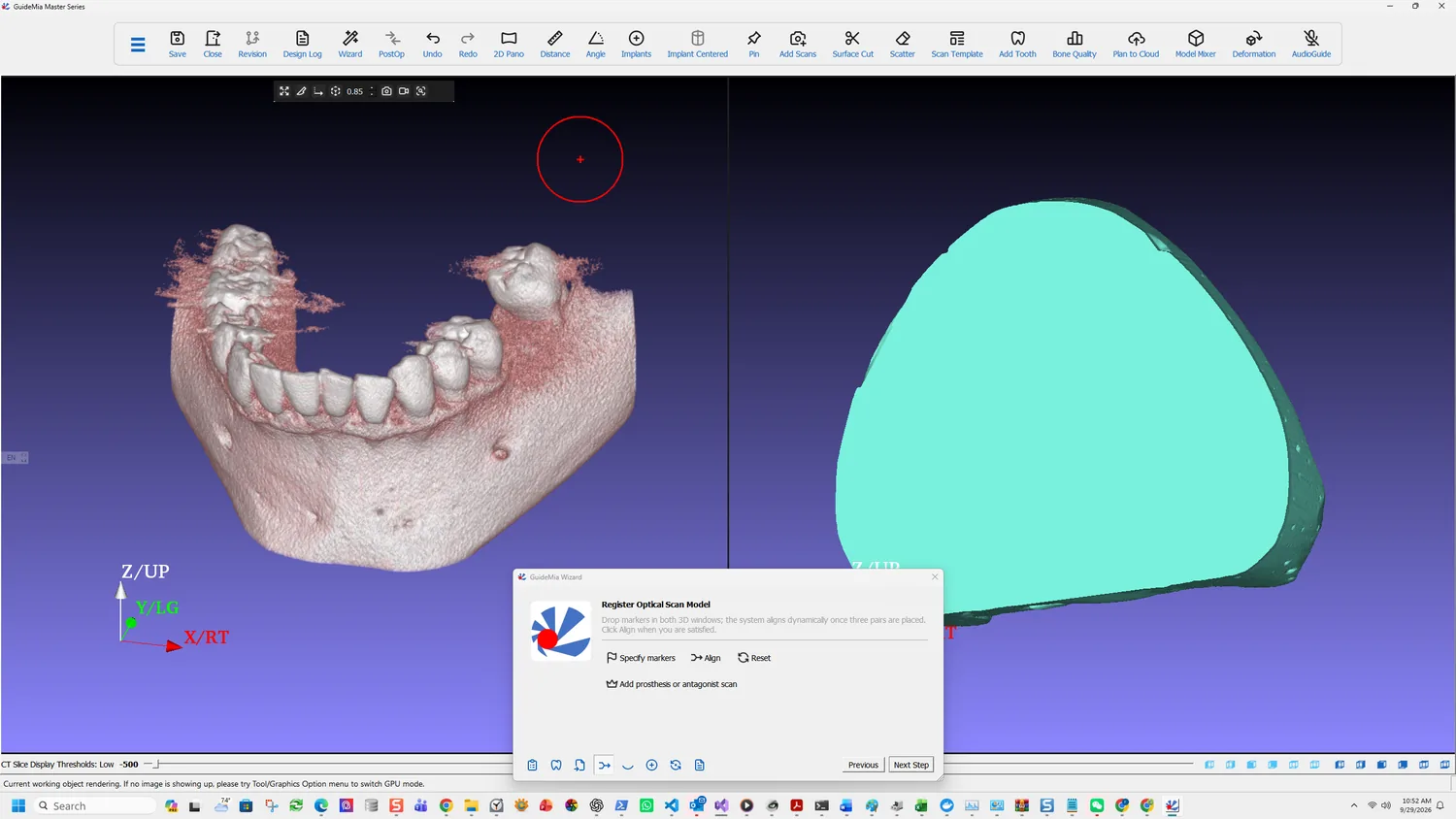
What you are looking at
After loading you have two models of the same arch that disagree about where they are in space. The CT-derived bone and teeth carry the scanner's coordinates; the optical scan carries its own. Neither is wrong, and nothing can be planned across both until they agree — which is the next step.
It is worth rotating both before registering. A scan with obvious defects — holes in the occlusal surfaces, a torn margin, a swallowed area of palate — is better fixed or re-taken now than discovered when the guide fails to cut. The software can repair a scan later, but the failure surfaces at guide design, several steps after the cause.
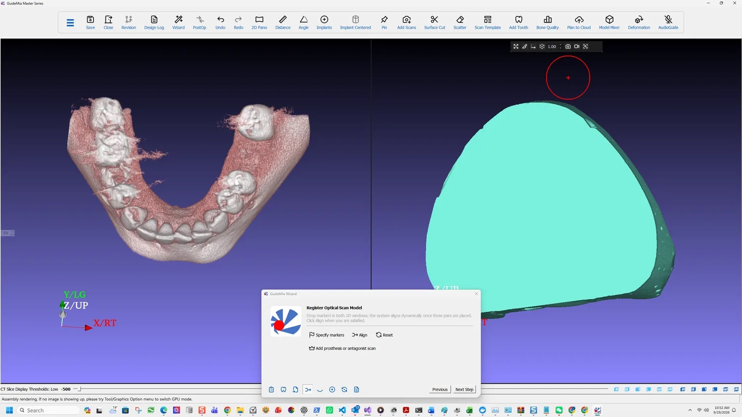
Turn it over first. The underside tells you whether the scan was trimmed cleanly and whether it is closed; a scan that is open where the guide will seat is the one that defeats Cut later.
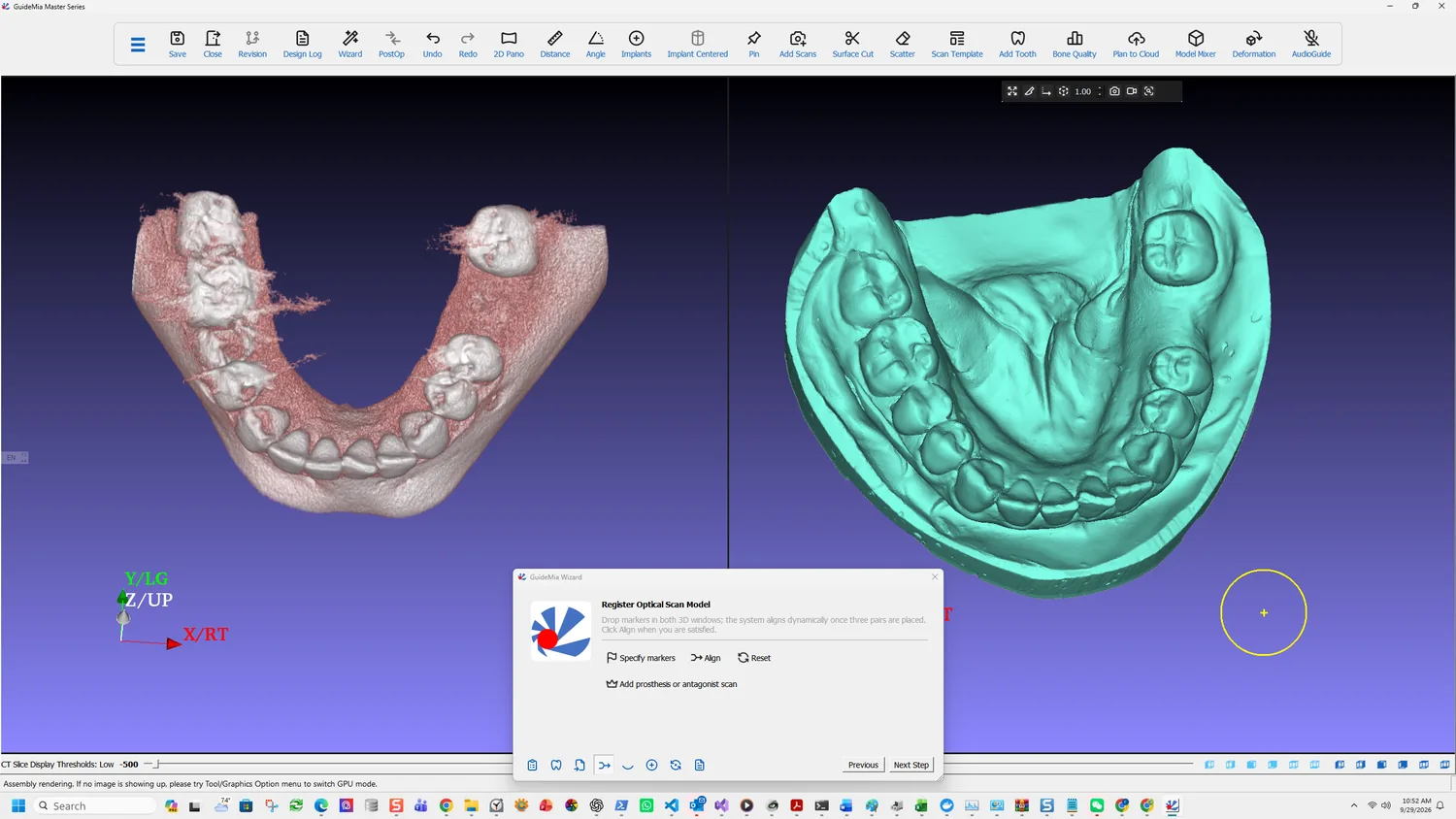
Then look at the occlusal surfaces, which are what the markers in the next step will be placed on and what the guide will rest on. You want unambiguous cusps and grooves. If the scan is soft there — smoothed over, or holed — the registration will be harder to place well and the fit will be harder to verify.
The full Load Case Data page carries the alternatives this case does not use:
- Radiographic Guide CT or optical — loads a radiographic guide's CBCT or optical scan. Two indications: a fully edentulous case, where the guide represents the gum surface and possible tooth arrangement; and a partially edentulous case where there is not enough natural tooth to register CBCT against a model scan, such as a patient with a long bridge.
- Impression CT or optical — loads an impression scan rather than a model scan. The model builder extracts the inner surface of the impression and turns it into a solid dental model, so an impression can substitute for a poured model.
- Denture design — loads a digital denture design, which can be registered against a radiographic guide or the gingiva model alone. GuideMia can then add abutment holes to it, or generate a prosthesis guide so the clinician can check occlusion and guide position before any drilling happens.
The Add Scans button on the main toolbar reaches the same loading functions without going through the Wizard.